

Transperineal Ultrasound (TPUS): An Exciting Technology
Key Messages
- An exciting use of transperineal ultrasound imaging is for the assessment of pelvic floor muscle function
- Recent research has highlighted the benefits of transperineal ultrasound for assessment of the male pelvic floor1-2
- Transperineal ultrasound is a valuable tool to use with patients before or after radical prostatectomy, prolapse, pelvic pain, obstructed defecation, or voiding dysfunction.
Real time 2D transperineal ultrasound imaging is emerging as an exciting technique for both pelvic floor muscle assessment and training. Far superior to the transabdominal approach, it’s a valuable tool to use with patients before or after radical prostatectomy, prolapse, pelvic pain, obstructed defecation, or voiding dysfunction.
Evolution Of Ultrasound Imaging For Physiotherapists
The use of real time 2D ultrasound imaging by physiotherapists has rapidly increased over the past few decades. It gained popularity for assessing and retraining the core abdominal muscles in patients with chronic low back pain, and is now used extensively across the profession to assess the function of many different muscle groups.
Transabdominal vs Transperineal Ultrasound
An exciting use of real time ultrasound imaging is for assessment of the pelvic floor muscles. This can be done via a transabdominal or transperineal approach.
The transabdominal approach gives a basic assessment of pelvic floor muscle function, but is limited by lack of bony landmarks, and poor anatomical detail. The transperineal approach is a far superior method of pelvic floor muscle assessment.
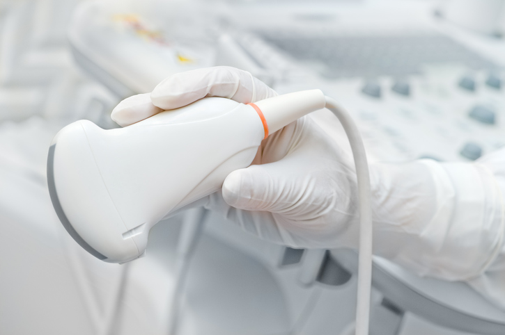
The ultrasound transducer is placed on the perineum, and a midline view of the pelvis is obtained. It allows simultaneous investigation of all the voluntary muscles of the pelvic floor, has proven reliability and validity, and is a powerful form of visual biofeedback1-3.

Fig 1: Patient positioning for transperineal ultrasound2
One limitation of ultrasound is that it doesn’t assess resting muscle tone. However, a skilled operator will be able to identify the poor motor control patterns associated with pelvic floor muscle overactivity, and can proceed to an internal exam for further investigation.
Transperineal Ultrasound In The Clinic
Transperineal ultrasound has only been used by physiotherapists in a clinical setting over the past few years. This is, in most part, due to the work of Professor Paul Hodges and Dr Ryan Stafford, research gurus at the University of Queensland. They have recently turned their attention to understanding the male pelvic floor and continence mechanism, and have been using transperineal ultrasound as their main assessment technique.
Using Transperineal Ultrasound For The Male Pelvic Floor
Historically, the male pelvic floor has been assessed via digital rectal exam. This is an appropriate way to assess levator ani function, however doesn’t give us information on the anterior urinary sphincter complex. The major advantage of the transperineal ultrasound image is that it allows simultaneous investigation of all the striated muscles contributing to continence. This is especially helpful when assessing men before or after radical prostatectomy.

Fig 2: Midsagittal view of the male pelvic floor, with ultrasound transducer on the perineum1
Applications Of Transperineal Ultrasound
Transperineal ultrasound has many useful applications, including:
- Before or after radical prostatectomy: TPUS allows visualization of all striated muscles involved in male continence
- Prolapse: TPUS allows pelvic floor muscle assessment during functional tasks such as coughing
- Pelvic pain: if patients are unable to tolerate an internal examination, TPUS is a great alternative for assessment
- Obstructed defecation: Visual biofeedback in TPUS enables re-education of defecation technique
- Voiding dysfunction: Visual biofeedback in TPUS enables re-education of voiding technique
- Patients preferring not to have an internal examination
Transperineal ultrasound imaging is a really exciting tool in our toolkit. It improves our clinical practice, and leads to improved patient outcomes especially for men undergoing radical prostatectomy.
References
1 Stafford, R. E., Ashton-Miller, J. A., Constantinou, C. E., & Hodges, P. W. (2013). A New Method to Quantify Male Pelvic Floor Displacement From 2D Transperineal Ultrasound Images. Urology, 81(3).
2 Stafford, R. E., Ashton-Miller, J. A., Constantinou, C. E., & Hodges, P. W. (2012). Novel Insight into the Dynamics of Male Pelvic Floor Contractions Through Transperineal Ultrasound Imaging. The Journal of Urology, 188, 1224-1230.
3 Constantinou, C. E. (2009). Dynamics of female pelvic floor function using urodynamics, ultrasound and magnetic resonance imaging (MRI). European Journal of Obstetrics, Gynecology & Reproductive Biology. 144(Suppl 1): S159-S165.
February 2017
Read some of our recent articles and education & health promotion
material on our News page.